41 compound microscope diagram without labels
Hot and Cold Packs: A Thermochemistry Activity | Carolina.com Diagram your hot or cold pack. Include labels to indicate sizes and quantities of materials used. List all materials and quantities needed to create your thermal pack. Explain the steps that you will follow to build your thermal pack. Describe the safety precautions you will use when creating and testing the thermal pack. Labelled Diagram of Compound Microscope - Biology Discussion The below mentioned article provides a labelled diagram of compound microscope. Part # 1. The Stand: The stand is made up of a heavy foot which carries a curved inclinable limb or arm bearing the body tube. The foot is generally horse shoe-shaped structure (Fig. 2) which rests on table top or any other surface on which the microscope in kept.
Parts of a Compound Microscope and Their Functions Compound microscope magnification is determined by multiplying the eyepiece and objective powers. When viewed through a 5X eyepiece with a 10X objective, an item is magnified 5 x 10=50 times. The magnification is 10 x 45 = 450 times when using a 10X eyepiece and a 45X objective. How to Use the Compound Microscope
Compound microscope diagram without labels
Parts of Stereo Microscope (Dissecting microscope) - labeled diagram ... Stereo microscopes (also called Dissecting microscope) are branched out from other light microscopes for the application of viewing "3D" objects. These include substantial specimens, such as insects, feathers, leaves, rocks, sand grains, gems, coins, and stamps, etc. Functionally, a stereo microscope is like a powerful magnifying glass. Labeling the Parts of the Microscope Labeling the Parts of the Microscope This activity has been designed for use in homes and schools. Each microscope layout (both blank and the version with answers) are available as PDF downloads. You can view a more in-depth review of each part of the microscope here. Download the Label the Parts of the Microscope PDF printable version here. Looking at the Structure of Cells in the Microscope A typical animal cell is 10–20 μm in diameter, which is about one-fifth the size of the smallest particle visible to the naked eye. It was not until good light microscopes became available in the early part of the nineteenth century that all plant and animal tissues were discovered to be aggregates of individual cells. This discovery, proposed as the cell doctrine by Schleiden and …
Compound microscope diagram without labels. Parts of Stereo Microscope (Dissecting microscope) – labeled diagram ... Compared to a compound microscope where the objectives attached to the nosepiece can be seen and identified individually (based on color bands and their respective labels), the objectives of a dissecting microscope are located in a cylindrical cone and, therefore, are not directly seen. For the stereo microscope that comes with multiple objective lens sets (fixed power style), the … PDF An Introduction to The Compound Microscope microscope in an upright position using both hands. **When carrying the microscope, place one hand on the base and the other hand around the arm. **DO NOT PLACE THE MICROSCOPE IN AN UPSIDE DOWN POSITION. PIECES WILL FALL OUT. **Keep microscope away from the edge of the bench, particularly when not in use. **Make sure power cords are out of the way. Diagram of a Compound Microscope - Biology Discussion A bright-field or compound microscope is primarily used to enlarge or magnify the image of the object that is being viewed, which can not otherwise be seen by the naked eye. Magnification may be defined as the degree of enlargement of the image of an object provided by the microscope. Pin on Science Activities with Microscopes - Pinterest Free worksheets for labeling parts of the microscope including a worksheet that is ... Worksheet identifying the parts of the compound light microscope.
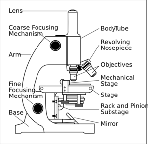
Binocular Microscope Anatomy - Parts and Functions with a Labeled Diagram Now, I will discuss the details anatomy of the light compound microscope with the labeled diagram. Why it is called binocular: because it has two ocular lenses or an eyepiece on the head that attaches to the objective lens, this ocular lens magnifies the image produced by the objective lens. Binocular microscope parts and functions Compound Microscope Parts – Labeled Diagram and their ... The term "compound" refers to the microscope having more than one lens. Basically, compound microscopes generate magnified images through an aligned pair of the objective lens and the ocular lens. In contrast, "simple microscopes" have only one convex lens and function more like glass magnifiers. Working Principle and Parts of a Compound Microscope (with Diagrams) It holds the stage, body tube, fine adjustment and coarse adjustment. 5. Body Tube: It is usually a vertical tube holding the eyepiece at the top and the revolving nosepiece with the objectives at the bottom. The length of the draw tube is called 'mechanical tube length' and is usually 140-180 mm (mostly 160 mm). 6. Label the microscope - Science Learning Hub In this interactive, you can label the different parts of a microscope. Use this with the Microscope parts activity to help students identify and label the main parts of a microscope and then describe their functions. Drag and drop the text labels onto the microscope diagram.
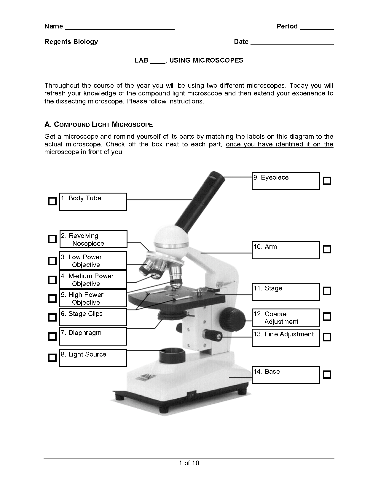
Compound Microscope Parts, Functions, and Labeled ... Nov 18, 2020 · Compound Microscope Definitions for Labels. Eyepiece (ocular lens) with or without Pointer: The part that is looked through at the top of the compound microscope. Eyepieces typically have a magnification between 5x & 30x. Monocular or Binocular Head: Structural support that holds & connects the eyepieces to the objective lenses. Compound Microscope: Definition, Diagram, Parts, Uses, Working ... - BYJUS The parts of a compound microscope can be classified into two: Non-optical parts Optical parts Non-optical parts Base The base is also known as the foot which is either U or horseshoe-shaped. It is a metallic structure that supports the entire microscope. Pillar The connection between the base and the arm are possible through the pillar. Arm Compound Microscope Parts Made Easy Not labeled in the above diagram but you can see it as a lightly glowing circle in the middle of the slide. (Above the reflection of the objective lens, which shows as a black circle). Illuminator - The illuminator provides the light for the microscope. It's usually a light bulb that's located underneath the stage. PDF Parts of a Microscope Printables - Homeschool Creations typical student microscope -other microscopes will vary) •Which part of the microscope rotates so another person can look through the eyepiece without needing to move the microscope ? the head •What is the magnification level on the eyepiece of a microscope?10x (see objective lens magnification to see how these work together)
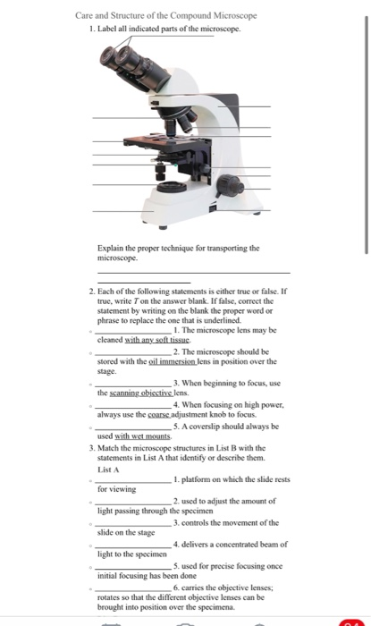
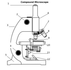
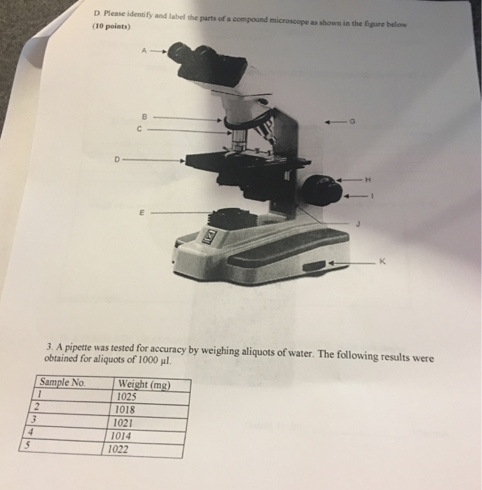
Care and Structure of the Compound Microscope 1. | Chegg.com
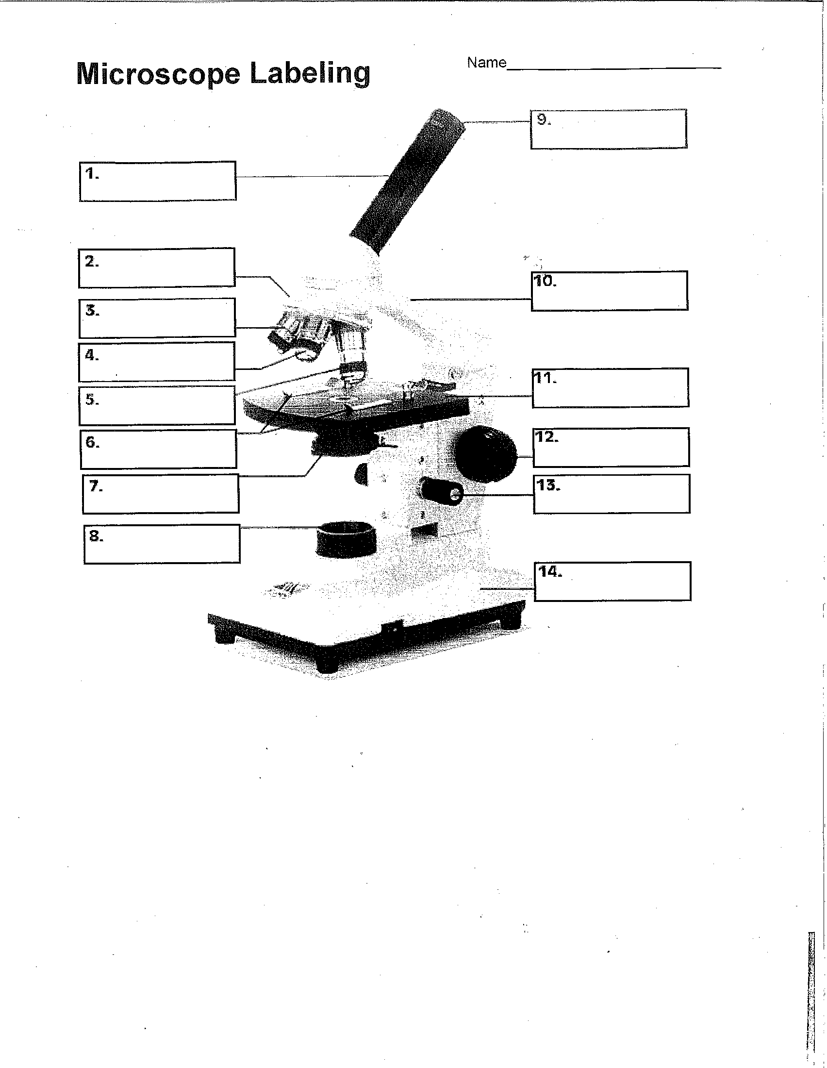
PDF Label compound microscope worksheet [clearBoth] [clearBoth] Microscope diagram without label After you've studied all the pieces of the composite microscope, it's time to put your brain to the test. Print an unmarked microscope chart and check that you can fill out all the blanks. [clearBoth] [clearBoth] Blank microscope diagram Next we have an empty microscope diagram.
Compound Microscope Clip Art at Clker.com - vector clip art online, royalty free & public domain
News Archives | Hollywood.com Travel through time by exploring Hollywood.com's entertainment news archives, with 30+ years of entertainment news content.

Microscope With Labels Clip Art at Clker.com - vector clip art online, royalty free & public domain
Inorganic Chemistry 4th edition, Catherine Housecroft Inorganic Chemistry 4th edition, Catherine Housecroft
30 Label The Parts Of A Compound Microscope - Labels Design Ideas 2020
PDF The Compound Light Microscope - teachers.wrdsb.ca The Compound Light Microscope TASK Refer to page 605 in your text to: 1. Name each of the structures described in the table to the right. 2. Match each structure to the letter in the diagram below. ** ALWAYS USE TWO HANDS TO CARRY A MICROSCOPE** Letter Structure Function joins body tube to base supports the entire microscope

Compound Microscope 3d Diagram - Micropedia
Two-Photon Excitation Microscopy for the Study of Living Cells … In a confocal microscope, a confocal pinhole is used to reject the out-of-focus background and produce an unblurred image which corresponds to a thin (<1 μm) “optical section.” This greatly reduces the limitation of imaging extended samples and enables high-resolution 3-D imaging. Two-photon excitation microscopy (a type of nonlinear microscopy, also known as multi …
Microscope With Labels Clip Art at Clker.com - vector clip art online, royalty free & public domain
Parts of a microscope with functions and labeled diagram Figure: Diagram of parts of a microscope There are three structural parts of the microscope i.e. head, base, and arm. Head - This is also known as the body. It carries the optical parts in the upper part of the microscope. Base - It acts as microscopes support. It also carries microscopic illuminators.

diagram of compound microscope - Brainly.in
Simple Microscope - Parts, Functions, Diagram and Labelling A compound microscope is also called a bright field microscope. It can provide magnification by up to 1,000 times. Stereo microscope/dissecting microscope - It can magnify objects by up to 300 times. It is used to visualize opaque objects that cannot be visualized using a compound microscope. Confocal microscope - It uses laser light to ...

Microscope - Science
Electron microscope - Wikipedia An electron microscope is a microscope that uses a beam of accelerated electrons as a source of illumination. As the wavelength of an electron can be up to 100,000 times shorter than that of visible light photons, electron microscopes have a higher resolving power than light microscopes and can reveal the structure of smaller objects.. Electron microscopes use shaped magnetic …

Diagram Of The Microscope - ClipArt Best
Compound Microscope- Definition, Labeled Diagram, Principle, Parts, Uses Alternatively, the magnification of the compound microscope is given by: m = D/ fo * L/fe where, D = Least distance of distinct vision (25 cm) L = Length of the microscope tube fo = Focal length of the objective lens fe = Focal length of the eye-piece lens Parts of a Compound Microscope Eyepiece And Body Tube.

Compound Microscope Diagram Hd - Micropedia
Microscope Parts and Functions - MICROSCOPEMASTER Microscope Parts and Functions With Labeled Diagram and Functions How does a Compound Microscope Work?. Before exploring microscope parts and functions, you should probably understand that the compound light microscope is more complicated than just a microscope with more than one lens.. First, the purpose of a microscope is to magnify a small object or to magnify the fine details of a larger ...
16 Best Images of Simple Microscope Labeling Worksheet - Compound Light Microscope Parts Blank ...
Compound Microscope - Types, Parts, Diagram, Functions and Uses It comes with a wide body and base. Its distinct parts include a condenser, illumination, focus lock, mechanical stage, and a revolving nosepiece which can hold up to five objectives. It usually has a binocular head, which makes long-term observation easy. Image 22: An example of a research compound microscope.

14 Best Images of Microscope Parts Function Worksheet Answers - Microscope Diagram Worksheet ...
Compound Microscope Parts, Diagram Definition, Application, Working ... Image: compound microscope labeled diagram | Image Source: microbiologynote.com. Head: It is located at the top portion of the microscope. It contains Eyepiece. ... It is not recommended to use oil immersion lenses without oil. Use of Compound microscope. Compound Microscopes used for blood analysis in pathological labs.

Compund Microscope Diagram - Diagram Resource Gallery
Parts of a Compound Microscope (And their Functions) List of Microscope Parts and their Functions. 1. Ocular Tubes (Monocular, Binocular & Trinocular) The ocular tubes, are to tubes that lead from the head of the microscope out to your eyes. On the end of the ocular tubes are usually interchangeable eyepieces (commonly 10X and 20X) that increase magnification.
Unit 2: Cell Biology - Mrs. Frump's Classes
How to Use a Compound Microscope: 11 Steps (with Pictures) To use a compound microscope, plug the microscope into a nearby outlet and prepare the slide with a cover slip or cover glass to protect the specimen. Next, place the slide on the center of the stage, over the glass hole, and use the two stage clips to secure the slide in place.
8 Best Images of Using A Microscope Worksheet - Compound Microscope Parts Blank, Microscope ...
Compound microscope - their parts and function - Microscopy4kids Compound microscopes have more than one lens to generate high magnification images of flat, thin specimens. 2. Eyepiece (10x) and Objective lenses (4x, 10x, 40x, 100x) are two major optical parts of a microscope. 3. Total magnification power is calculated by multiplying the magnification of the eyepiece and objective lens. 4.

Compound Microscope Drawing - Micropedia
Label Microscope Diagram - EnchantedLearning.com Using the terms listed below, label the microscope diagram. arm - this attaches the eyepiece and body tube to the base. base - this supports the microscope. body tube - the tube that supports the eyepiece. coarse focus adjustment - a knob that makes large adjustments to the focus. diaphragm - an adjustable opening under the stage, allowing ...
16 Best Images of Simple Microscope Labeling Worksheet - Compound Light Microscope Parts Blank ...
Fluorescence - Wikipedia Fluorescence is the emission of light by a substance that has absorbed light or other electromagnetic radiation.It is a form of luminescence.In most cases, the emitted light has a longer wavelength, and therefore a lower photon energy, than the absorbed radiation.A perceptible example of fluorescence occurs when the absorbed radiation is in the ultraviolet …
Solved: Label The Parts Of The Microscope Below And Descri... | Chegg.com
Microscope Labeling - The Biology Corner Students label the parts of the microscope in this photo of a basic laboratory light microscope. Can be used for practice or as a quiz.
Post a Comment for "41 compound microscope diagram without labels"