40 fluorescent labels and light microscopy
Different Ways to Add Fluorescent Labels - Thermo Fisher Scientific Using fluorescence provides greater contrast compared to viewing your samples with brightfield microscopy alone. Labeling various targets with separate fluorescent colors allows you to visualize different structures or proteins within a cell in the same experiment. Fluorescent labeling of abundant reactive entities (FLARE) for cleared ... Fluorescence microscopy is a vital tool in biomedical research but faces considerable challenges in achieving uniform or bright labeling. For instance, fluorescent proteins are limited to model ...
› articles › s41592/018/0219-4Expansion microscopy: principles and uses in biological ... Dec 20, 2018 · After gelation, digestion, and expansion, the fluorescence of the antibody labels or fluorescent proteins is largely retained for many genetically encoded fluorophore and small-molecule dyes (Fig ...
Fluorescent labels and light microscopy
Fluorescent Dyes Types, Vs Proteins, Applications Etc. Fluorescent dyes (also known as fluorophores/reactive dyes) may simply be described as molecules (non-protein in nature) that, in microscopy, achieve their function by absorbing light at a given wavelength and re-emitting it at a longer wavelength. This produces fluorescence of different colors that can be visualized and analyzed. Fluorescence Microscopy - PMC - PubMed Central (PMC) Fluorescence Light Separation. The goal of the fluorescence microscope is to separate emitted light (dim) from excitation light (bright) (Fig. 2); fluorescence indicators with large Stokes shifts are advantageous for this. The separation of the light is generally achieved with optical filters and the key to successful imaging is their selection ... Light Microscope- Definition, Principle, Types, Parts, Labeled Diagram ... A light microscope is a biology laboratory instrument or tool, that uses visible light to detect and magnify very small objects and enlarge them. They use lenses to focus light on the specimen, magnifying it thus producing an image. The specimen is normally placed close to the microscopic lens.
Fluorescent labels and light microscopy. › techniques › multi-photonMultiphoton Microscopy | Nikon’s MicroscopyU In the descanned geometry, the emitted light (illustrated in blue) returns along the same path as the excitation light, striking the scanning mirrors before passing through the confocal pinhole to the detector (Figure 4(a)). In confocal microscopy this geometry must be utilized to eliminate the detection of out-of-focus emission. Imaging Flies by Fluorescence Microscopy: Principles, Technologies, and ... The development of fluorescent labels and powerful imaging technologies in the last two decades has revolutionized the field of fluorescence microscopy, which is now widely used in diverse scientific fields from biology to biomedical and materials science. ... has brought about the era of fluorescence light microscopy. The first fluorescence ... Fluorescence Microscopy vs. Light Microscopy - News-Medical.net This means that fluorescent microscopy uses reflected rather than transmitted light. For example, a commonly used label is green fluorescent protein (GFP), which is excited with blue light and... Fluorescence Live Cell Imaging - PMC - PubMed Central (PMC) Fluorescent protein tags, live cell dyes, and other methods to fluorescently label proteins of interest provide a range of tools to investigate virtually any cellular process under the microscope. The two main experimental challenges in collecting meaningful live cell microscopy data are to minimize photodamage while retaining a useful signal ...
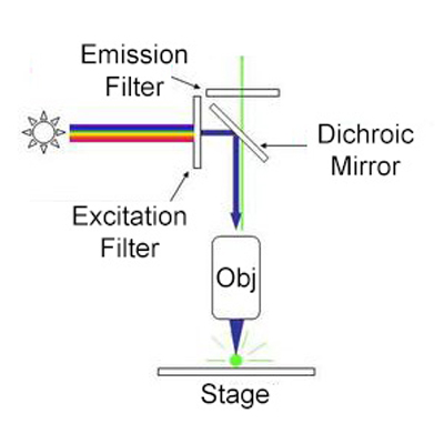
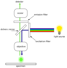
Fluorescence Microscopy & Cell Imaging | Research | UNM Cancer Center The Fluorescence Microscopy and Cell Imaging Shared Resource aids basic and physician researchers to image samples and publish high profile articles that: Elucidate cell and molecular mechanisms of cancer, immunologic, infectious, metabolic, neurologic and vascular diseases. Evaluate therapeutic efficacy in cells and patient samples. Fluorescence microscope - Wikipedia Typical components of a fluorescence microscope are a light source ( xenon arc lamp or mercury-vapor lamp are common; more advanced forms are high-power LEDs and lasers ), the excitation filter, the dichroic mirror (or dichroic beamsplitter ), and the emission filter (see figure below). In Silico Labeling: Predicting Fluorescent Labels in Unlabeled Images Microscopy is a central method in life sciences. Many popular methods, such as antibody labeling, are used to add physical fluorescent labels to specific cellular constituents. ... (ISL), reliably predicts some fluorescent labels from transmitted-light images of unlabeled fixed or live biological samples. ISL predicts a range of labels, such as ... Fluorescence Microscopy - Explanation and Labelled Images Fluorescence microscopy uses a high-intensity light source that excites a fluorescent molecule called a fluorophore in the sample observed. The samples are labeled with fluorophore where they absorb the high-intensity light from the source and emit a lower energy light of longer wavelength.
Fluorescent Dyes | Science Lab | Leica Microsystems In fluorescence microscopy there are two ways to visualize your protein of interest. Either with the help of an intrinsic fluorescent signal - by genetically linking a fluorescent protein to a target protein - or with the help of fluorescently labeled antibodies that bind specifically to a protein of interest. Label-free prediction of three-dimensional fluorescence images from ... We present a label-free method for predicting three-dimensional fluorescence directly from transmitted-light images and demonstrate that it can be used to generate multi-structure, integrated... en.wikipedia.org › wiki › FluorophoreFluorophore - Wikipedia Fluorophore molecules could be either utilized alone, or serve as a fluorescent motif of a functional system. Based on molecular complexity and synthetic methods, fluorophore molecules could be generally classified into four categories: proteins and peptides, small organic compounds, synthetic oligomers and polymers, and multi-component systems. Fluorescence Microscopy Buyer's Guide - Biocompare Seeing is believing, and for many biologists, what they want to see is fluorescent labels through a microscope. Fluorescence microscopy, which makes it possible to visualize fluorescent proteins or dyes at the cellular and subcellular level, has become a workhorse of modern biology.. The concept of fluorescence, in which a molecule absorbs light of one wavelength and then emits light of a ...
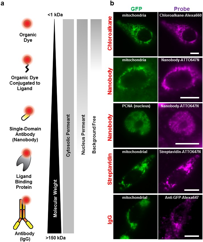
Labeling proteins inside living cells using external ...
Fluorescence microscopy: established and emerging methods ... - PubMed The primary concern in all forms of microscopy is the generation of contrast; for fluorescence microscopy contrast can be thought of as the difference in intensity between the cell and background, the signal-to-noise ratio. High information-content images can be formed by enhancing the signal, suppressing the noise, or both.

Feature-rich covalent stains for super-resolution and cleared ...
› articles › s41467/022/33157-4Payload distribution and capacity of mRNA lipid nanoparticles Sep 23, 2022 · a Species of interest in LNP formulations include the mRNA-loaded LNPs, empty LNPs, and free mRNAs. Three fluorescent tags were used for the single-particle fluorescence detection and species ...

Label-free prediction of three-dimensional fluorescence ...
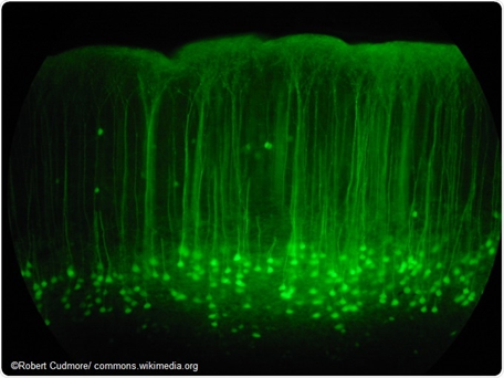
Fluorescent Microscopy Fluorescent microscopy is often used to image specific features of small specimens such as microbes. It is also used to visually enhance 3-D features at small scales. This can be accomplished by attaching fluorescent tags to anti-bodies that in turn attach to targeted features, or by staining in a less specific manner.

Fluorescent protein tags | Proteintech Group
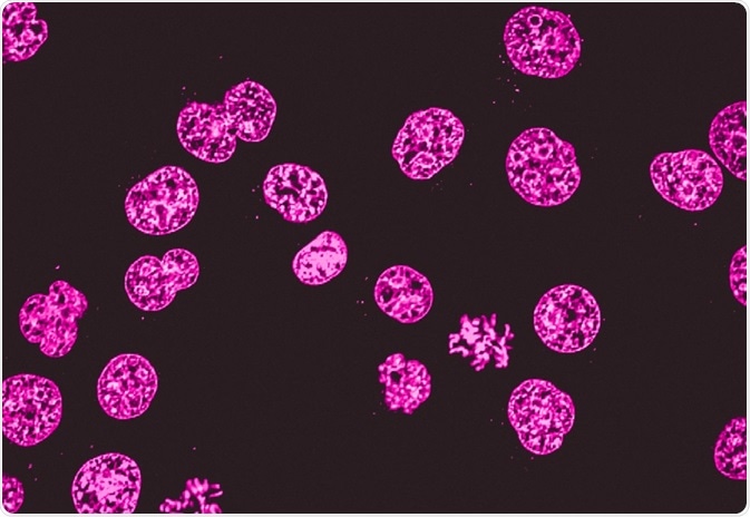
Novel Fluorescent Label Shines a Light on DNA Structure in Cancer Cells Novel Fluorescent Label Shines a Light on DNA Structure in Cancer Cells March 7, 2022 Researchers have developed a new fluorescent label that gives a clearer picture of how DNA architecture is...

VIPER is a genetically encoded peptide tag for fluorescence ...
Fluorescent Labeling - What You Should Know - PromoCell Fluorescence microscopy allows the identification of cells and cellular components and the monitoring of cell physiology with high specificity. Fluorescence microscopy separates emitted light from excitation light using optical filters. The use of two indicators also allows the simultaneous observation of different biomolecules at the same time.
IJMS | Free Full-Text | Pyrene Excimer-Based Fluorescent ...
en.wikipedia.org › wiki › FluorescenceFluorescence - Wikipedia Fluorescence is the emission of light by a substance that has absorbed light or other electromagnetic radiation.It is a form of luminescence.In most cases, the emitted light has a longer wavelength, and therefore a lower photon energy, than the absorbed radiation.
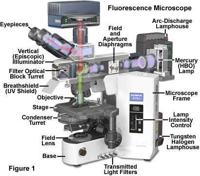
Fluorescence Microscopy - Anatomy of the Fluorescence ...
Light Microscopy Techniques in Virology: An Overview - News-Medical.net Then a suitable antibody with a fluorescent label added, with the unbound antibody removed through a series of washes. The cells can then be studied by utilizing a suitable light microscopy method. Direct immunolabeling involves the direct binding of a fluorescent label to a viral antigen.

Fluorescent Labeling - What You Should Know - PromoCell
Introduction to Fluorescence Microscopy • iBiology In this introductory lecture on light microscopy, Dr. Nico Stuurman describes the principles and properties of fluorescence microscopy. Skip to primary navigation; ... 00:01:04;23 so we label microtubules with the fluorescent dye 00:01:08;13 and we have part of the cell labeled in red, 00:01:11;25 and that part is actually the uh, nucleus
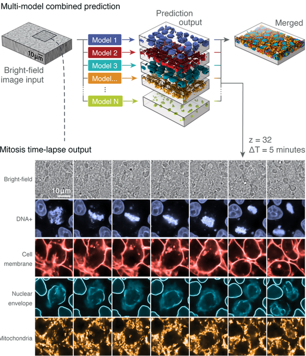
Label-free Determination - ALLEN CELL EXPLORER
en.wikipedia.org › wiki › ImmunofluorescenceImmunofluorescence - Wikipedia Immunofluorescence is a technique used for light microscopy with a fluorescence microscope and is used primarily on microbiological samples. This technique uses the specificity of antibodies to their antigen to target fluorescent dyes to specific biomolecule targets within a cell, and therefore allows visualization of the distribution of the target molecule through the sample.
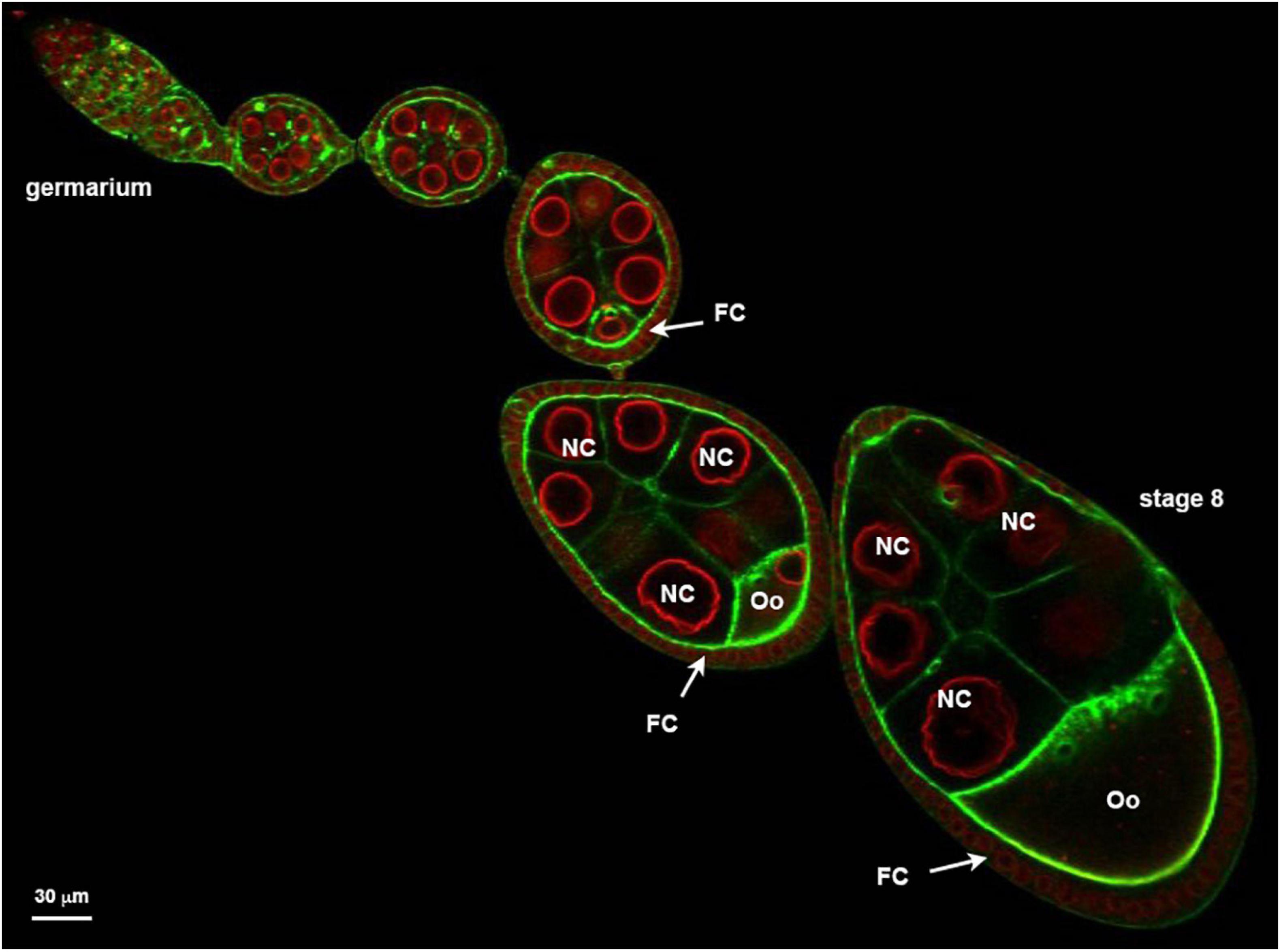
The Biological bulletin. Biology; Zoology; Biology; Marine ...
Fluorescence Microscopy - an overview | ScienceDirect Topics Fluorescence microscopy is a technique whereby fluorescent substances are examined in a microscope. It has a number of advantages over other forms of microscopy, offering high sensitivity and specificity. In fluorescence microscopy, the specimen is illuminated (excited) with light of a relatively short wavelength, usually blue or ultraviolet (UV).
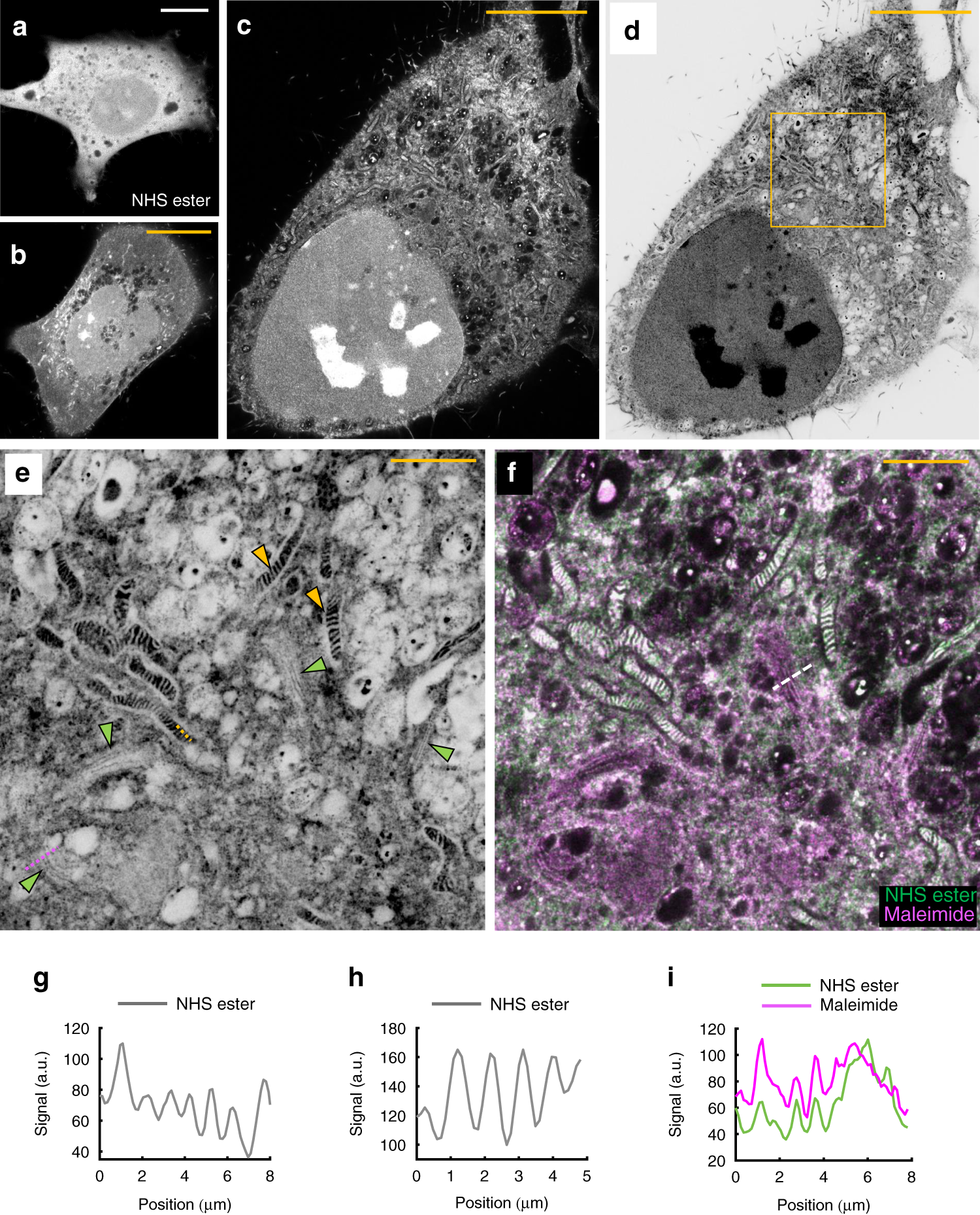
Light microscopy of proteins in their ultrastructural context ...
Fluorescent tag - Wikipedia S. cerevisiae septins revealed with fluorescent microscopy utilizing fluorescent labeling In molecular biology and biotechnology, a fluorescent tag, also known as a fluorescent label or fluorescent probe, is a molecule that is attached chemically to aid in the detection of a biomolecule such as a protein, antibody, or amino acid.
Fluorescence Microscopy - Explanation and Labelled Images ...
Fluorescence Microscope: Principle, Types, Applications Fluorescence microscopy is a light microscope that works on the principle of fluorescence. A substance is said to be fluorescent when it absorbs the energy of invisible shorter wavelength radiation (such as UV light) and emits longer wavelength radiation of visible light (such as green or red light).
FluoroNanogold™ combined fluorescent and gold nanoparticle ...
Fluorescence Imaging - Teledyne Photometrics Fluorescent molecules (known as fluorophores) are used to label samples, and fluorophores are available that emit light in virtually any color. In a fluorescent microscope, a sample is labeled with a fluorophore, and then a bright light ( excitation light) is used to illuminate the sample, which gives off fluorescence ( emission light ).

Super-resolution Microscopy – CENT – Center for Enhanced ...
Introduction to Fluorescence Microscopy | Nikon's MicroscopyU It is important to note that fluorescence is the only mode in optical microscopy where the specimen, subsequent to excitation, produces its own light. The emitted light re-radiates spherically in all directions, regardless of the excitation light source direction.
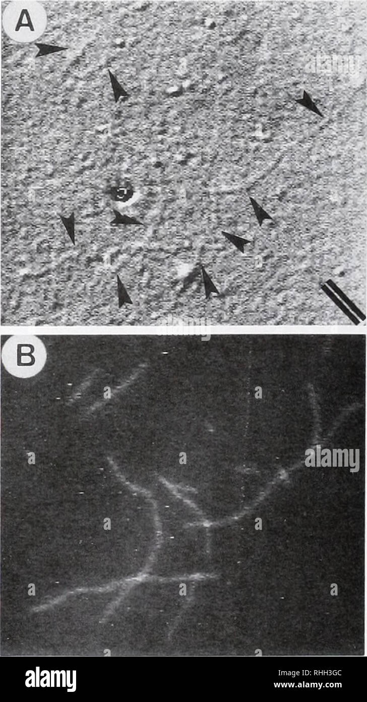
The Biological bulletin. Biology; Zoology; Biology; Marine ...
› techniques › fluorescenceIntroduction to Fluorescent Proteins | Nikon’s MicroscopyU The brightness and fluorescence emission spectrum of enhanced yellow fluorescent protein combine to make this probe an excellent candidate for multicolor imaging experiments in fluorescence microscopy. Enhanced yellow fluorescent protein is also useful for energy transfer experiments when paired with enhanced cyan fluorescent protein (ECFP) or ...
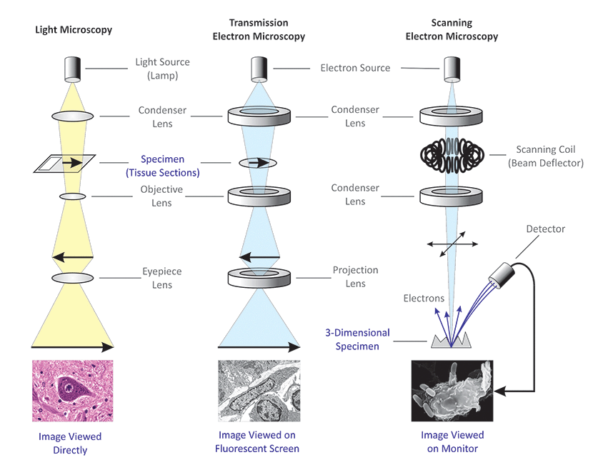
Differences between Light Microscope and Electron Microscope
In Silico Labeling: Predicting Fluorescent Labels in Unlabeled ... - Cell The z-stacks of transmitted-light microscopy images were acquired with different methods for enhancing contrast in unlabeled images. Several different fluorescent labels were used to generate fluorescence images and were varied between training examples; the checkerboard images indicate fluorescent labels that were not acquired for a given example.
Fluorescence microscope - Wikipedia
Labeling Proteins For Single Molecule Imaging - Teledyne Photometrics A fluorescent signal allows a protein's precise location to be determined as well as its behavior to be observed. This can then be studied in relation to other fluorescently tagged proteins. In this way, researchers are able to use fluorescence microscopy to visualize underlying biological processes.

Fluorescent Dyes | Science Lab | Leica Microsystems
Important biomedical microscopy technique can now image ... - ScienceDaily The light needed to image fluorescent labels can be damaging and even deadly to delicate biological samples such as brain tissue or animal embryos used to study development and disease processes ...
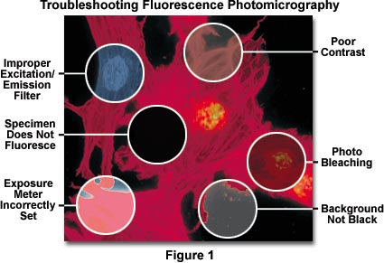
Fluorescence Microscopy Errors | Olympus LS
Light Microscope- Definition, Principle, Types, Parts, Labeled Diagram ... A light microscope is a biology laboratory instrument or tool, that uses visible light to detect and magnify very small objects and enlarge them. They use lenses to focus light on the specimen, magnifying it thus producing an image. The specimen is normally placed close to the microscopic lens.
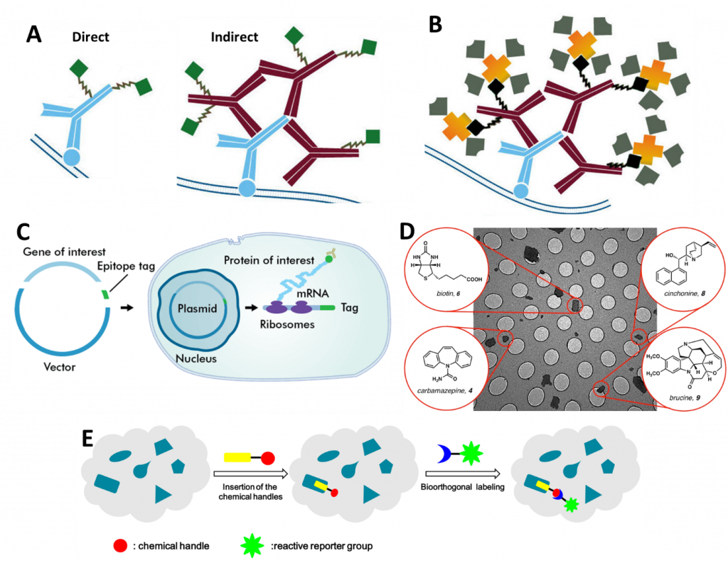
Protein Labeling with Fluorescent Probes - Theory and Methods
Fluorescence Microscopy - PMC - PubMed Central (PMC) Fluorescence Light Separation. The goal of the fluorescence microscope is to separate emitted light (dim) from excitation light (bright) (Fig. 2); fluorescence indicators with large Stokes shifts are advantageous for this. The separation of the light is generally achieved with optical filters and the key to successful imaging is their selection ...
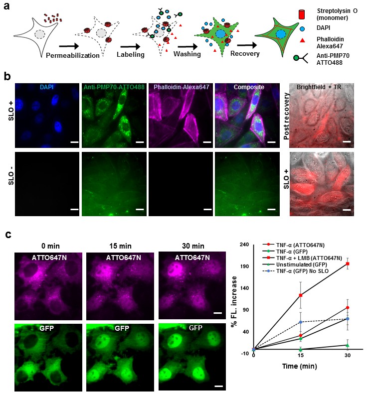
Labeling proteins inside living cells using external ...
Fluorescent Dyes Types, Vs Proteins, Applications Etc. Fluorescent dyes (also known as fluorophores/reactive dyes) may simply be described as molecules (non-protein in nature) that, in microscopy, achieve their function by absorbing light at a given wavelength and re-emitting it at a longer wavelength. This produces fluorescence of different colors that can be visualized and analyzed.
In Silico Labeling: Predicting Fluorescent Labels in ...

Fluorescent Labelling, FRET & Light Sheet Microscopy – Ben Sutcliffe
Frontiers | GFP-Tagged Protein Detection by Electron ...
Labeling Proteins For Single Molecule Imaging

Light microscopes — Science Learning Hub

Fluorescent tag - Wikipedia

Immunofluorescence Microscopy. Immunofluorescence Microscopy ...
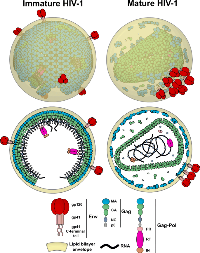
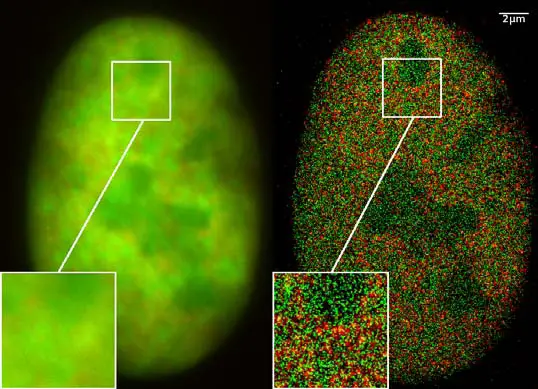
Super-resolution fluorescence microscopy studies of human ...
Hyperspectral multiphoton microscopy for in vivo ...
Fluorescence Microscopy vs. Light Microscopy
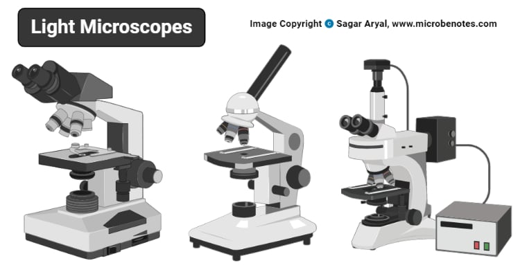
Light Microscope- Definition, Principle, Types, Parts ...
Correlative fluorescence microscopy, transmission electron ...
Fluorescent Dyes in Microscopy - Types, Vs Proteins ...

Practical Fluorescence Reconstruction Microscopy for High ...

VIPER is a genetically encoded peptide tag for fluorescence ...

An Overview of Fluorescence Microscopy - Biotium

Bacterial Vivisection: How Fluorescence-Based Imaging ...

Label-free imaging tool pipeline and application using 3D ...

Multi-label in vivo STED microscopy by parallelized switching ...
Post a Comment for "40 fluorescent labels and light microscopy"